Fig. 1.
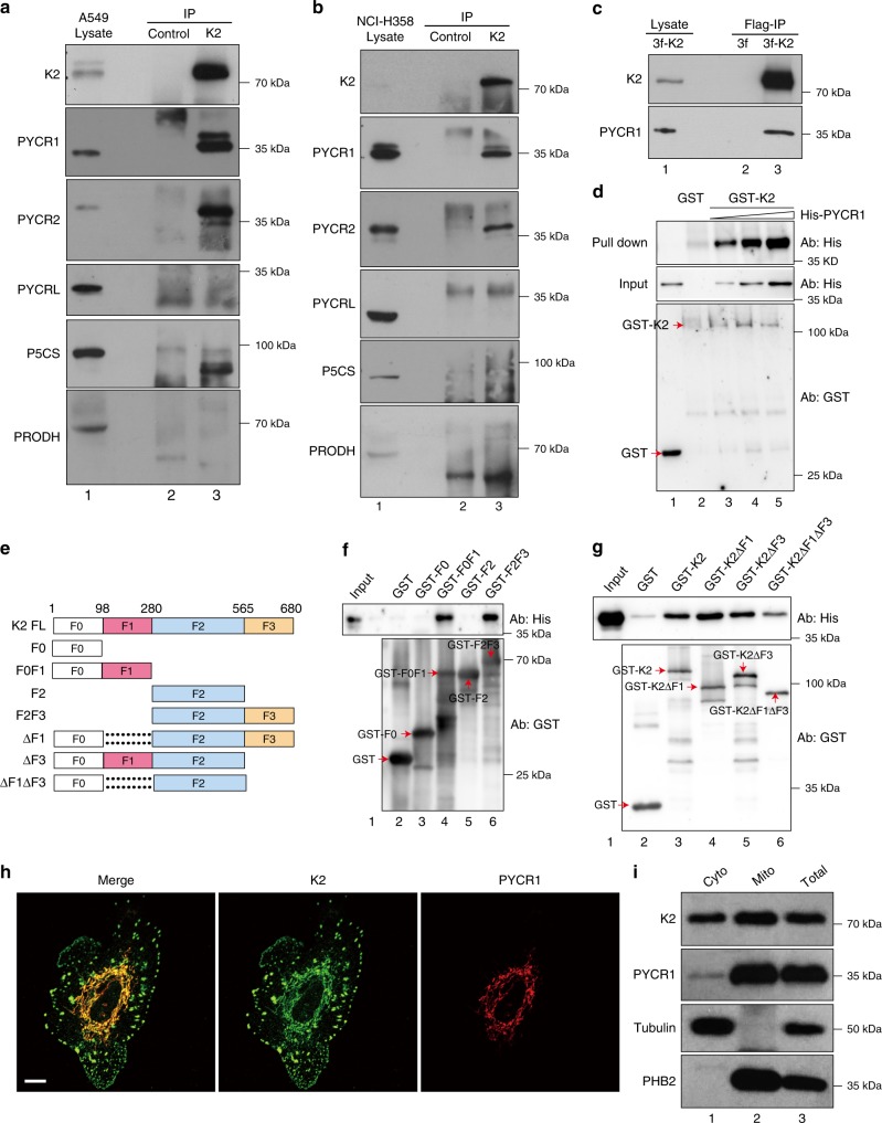
Kindlin-2 interacts with PYCR1 and colocalizes with PYCR1 in mitochondria. a, b Co-IP of PYCR1 with kindlin-2. Human A549 (a) or HCI-H358 (b) cells were analyzed by IP with monoclonal anti-kindlin-2 antibody or irrelevant mouse IgG (as a control) as described in the Methods. The cell lysates (lane 1), control IgG (lane 2), and anti-kindlin-2 immunoprecipitates (lane 3) were analyzed by western blotting with antibodies as indicated. c Co-IP of PYCR1 with FLAG-kindlin-2. Kindlin-2 KO A549 cells were infected with lentiviral vector encoding 3xFLAG-tagged kindlin-2 (3fl-K2) or control lentiviral vector lacking kindlin-2 sequence (3fl). The 3fl-K2 and control 3fl infectants were analyzed by IP with anti-FLAG antibody. The 3fl-K2 cell lysates (lane 1) and IP samples from 3fl (lane 2) or 3fl-K2 (lane 3) cells were analyzed by western blotting with kindlin-2 or PYCR1 antibodies. d GST-kindlin-2 bound to glutathione-Sepharose beads were incubated with increased concentrations (lane 2, 0 μg ml−1; lane 3, 10 μg ml−1; lane 4, 20 μg ml−1; lane 5, 40 μg ml−1) of purified His-tagged PYCR1. GST-kindlin-2 fusion protein pulldown was analyzed as described in the Methods. The sample in lane 1 was prepared as that in lane 4, except GST-kindlin-2 was replaced with GST. e–g GST-tagged full-length or mutant forms of kindlin-2 (illustrated in e) or GST alone (as a negative control) were used to pull down His-tagged PYCR1 as described in the Methods. The inputs, GST and GST-kindlin-2 fusion protein pulldowns, were analyzed by western blotting with antibodies against His and GST, respectively. h A549 cells were plated on fibronectin-coated coverslips and dually stained with mouse monoclonal anti-kindlin-2 and rabbit polyclonal anti-PYCR1 antibodies. The primary antibodies were detected with Alexa Fluor 488-conjugated anti-mouse IgG or Alexa Fluor 647-conjugated anti-rabbit IgG secondary antibodies. Scale bar = 10 μm. i The cytosolic fraction (Cyto, lane 1), mitochondrial fraction (Mito, lane 2), and total cell lysates (Total, lane 3) were analyzed by western blotting with antibodies to kindlin-2, PYCR1, tubulin, and prohibitin-2 (PHB2) as indicated

