Fig. 10.
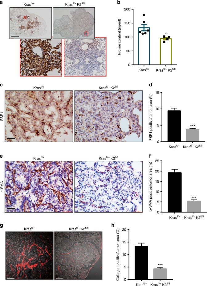
Ablation of kindlin-2 in lung adenocarcinoma substantially reduces PYCR1 and proline levels, and fibrosis in vivo. a–h The lung of the mice (as indicated in the figure) was administrated with Ad-Cre and analyzed 16 weeks later. Sections of the lung tissues from the mice (as specified in the figure) were analyzed by immunostaining with anti-PYCR1 antibody (a). Scale bar = 300 μm. The proline levels in the lung tissues were analyzed as described in the Methods (b, Kras fl/+ group n = 6; Kras fl/+; K2fl/fl group n = 4). Sections of the lung tissues from the mice (as specified in the figure) were analyzed by immunostaining with an antibody for fibroblast-specific protein 1 (FSP1), a marker of fibroblasts (c). Scale bar = 20 μm. (d) The percentages of FSP1-positive areas among total tumor areas were quantified (at least 25 fields from 4 mice were counted). Sections of the lung tissues from the mice (as specified in the figure) were analyzed by immunostaining with an antibody for myofibroblast marker α-SMA (e). Scale bar = 20 μm. f The percentages of α-SMA-positive areas among total tumor areas were quantified (at least 35 fields from 4 mice were counted). Sections of the lung tissues from the mice (as indicated in the figure) in which collagen fibers were analyzed by second harmonic generation (SHG) with multiphoton microscopy are shown (g). Scale bar in g, 100 μm. The percentages of collagen matrix-positive areas among total tumor areas were quantified (h, at least 30 fields from 4 mice were counted). Data in b, d, f, h represent mean ± SEM. Statistical significance was calculated using two-tailed unpaired Student’s t-test, *P < 0.05; ***P < 0.001

