Fig. 5.
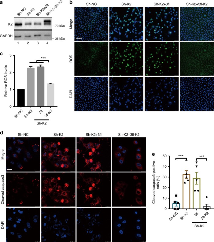
Depletion of kindlin-2 increases ROS production and apoptosis. a–c A549 cells were infected with K2 shRNA (Sh-K2) lentivirus or control lentivirus (Sh-NC). Two days after the infection, the cells were infected with lentiviral expression vectors encoding 3xFLAG-tagged kindlin-2 (3fl-K2) or 3xFLAG empty vector (3fl). Three days after the lentiviral infection, the cells were analyzed by western blotting with antibodies recognizing K2 or GAPDH (as a loading control) (a). The levels of ROS were analyzed using DHE fluorescence probe as described in the Methods (b). Scale bar, 75 μm. The mean fluorescence intensities (MFI) were calculated using the Image-pro plus software from two different experiments (c, at least n = 15 field per group were counted from at least three independent experiments). d The cells (as specified in the figure) were immunofluorescently stained with DAPI and anti-cleaved caspase-3 antibody. Scale bar = 25 μm. The percentages of cleaved caspase-3-positive cells were quantified (e) as described in the Methods. Replicates (n = 5) from one representative experiment are shown. Data in c and e are presented as mean ± SEM using one-way ANOVA with Tukey–Kramer post-hoc analysis, *P < 0.05; ***p < 0.001

