Fig. 2.
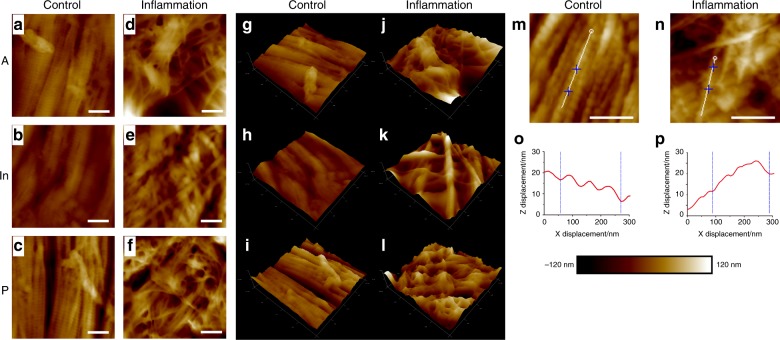
AFM mapping scanning images of TMJ discs. a–c Representative morphologic features of discs from the control group. The collagen fibrils exhibited good arrangement with clear light and dark banding of the D-period. The arrangement of anterior bands and intermediate zones were similar, and the posterior band was slightly loosened but still in good order. d–f Representative morphologic features of the discs from the inflamed group. The banding of collagen fibrils was diminished, and many single fibrils were observed. The spatial arrangement of collagen bundles was disordered and non-directional. g–l Three-dimensional reconstruction of a–f images, respectively. The inflammation group presented porous and undulated structures compared with the control group. Representative images of collagen single micro-fibrils from control and inflamed TMJ discs, respectively. o, p x–z directional curve of single collagen micro-fibril corresponding to the white line in m and n. The topographic structure exhibited good repeating D-periods in control discs and degraded performance in inflamed discs. Scale bar: 400 nm

