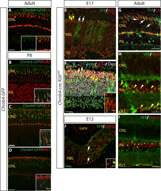
Figure 1.
Chrnb4-GFP and Chrnb4-cre expression in developing and adult cone photoreceptor cells. (A) Immunostaining of adult (6 week old) Chrnb4-GFP retinas. Chrnb4-GFP expression co-labels with the expression of cone marker CA in the ONL. (B–D) Immunostaining of P8 Chrnb4-GFP retinas. Chrnb4-GFP expression co-labels with the expression of cone markers RxRγ (B) and CA (C) but not with the expression of rod marker RHO (D). See Supplementary Figs 3 and 4 for the separate channels of the data shown in the merged images in A & B. (E,F) Immunostaining on E17 Chrnb4-cre; R26YFP retinas using an anti-GFP antibody to amplify the YFP signal intensity. YFP expression is detected in some RxRγ+ve cones (white arrows), while most cones remain YFP-ve (E, yellow arrows). YFP expression is also seen in some OTX2-expressing photoreceptor progenitors (F, white arrows). Yellow arrows indicate OTX2+ve cells that are not YFP+ve. OTX2+ve RPE cells do not express YFP (blue arrows). (G–I) Immunostaining on adult (6 weeks old) Chrnb4-cre; R26YFP retinas. YFP expression was detected in CA+ve cones (G, white arrows) and in a few inner retinal cells including some PAX6+ve cells (H, white arrowheads). Most Pax6+ve cells in the inner retina remained YFP-ve (H, yellow arrowheads). CHX10+ve bipolar cells were mainly YFP-ve (I, asterisks). (J) Immunostaining on E12 Chrnb4-cre; R26YFP retinas was also performed. YFP expression was detected in a subset of CHX10+ve RPCs (white arrows). CA: cone arrestin. RxRγ: retinoid x receptor gamma. RHO: rhodopsin. RPE: retinal pigment epithelium. ONL: outer nuclear layer. INL: inner nuclear layer. GCL: ganglion cell layer. RPC: retinal progenitor cells. Scale bars: 30 μm. Scale bars of insets: 10 μm.

