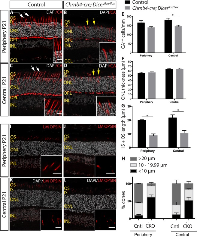
Figure 3.
Cone abnormalities by P21 in Dicer CKO mice. (A–D) Immunostaining for CA of P21 control (A,C) and Dicer CKO (B,D) peripheral and central retinas. White arrows indicate normal wild type segments. Yellow arrows indicate abnormal short segments (E) Quantification of CA+ve cone photoreceptors. (F) Measurement of the ONL thickness. (G) Average length of CA+ve segments (IS + OS). (H) Graph showing the percentage of cones displaying long (>20 μm), medium (10–19.99 μm) and short segments (<10 μm) in control and Dicer CKO peripheral and central retinas. (I–L) Immunostaining for the cone outer segment marker LM OPSIN. OS: outer segments. IS: inner segments. ONL: outer nuclear layer. INL: inner nuclear layer. OPL: outer plexiform layer. GCL: ganglion cell layer. CA: cone arrestin. *Indicates p-value < 0.05. Mann-Whitney non parametric test was used. Error bars indicate standard error of the mean. Scale bar: 30 μm. Scale bar of insets: 10 μm.

