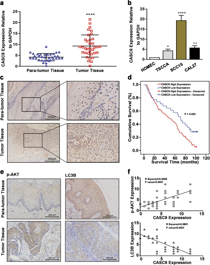
Fig. 1. CASC9 is highly expressed in OSCC tissues and cells.
a RT-qPCR results showed that CASC9 expression was significantly increased in OSCC tissues compared with paired adjacent tissues (N = 35). b RT-qPCR results showed that CASC9 expression was significantly increased in TSCCA, SCC15 and CAL27 OSCC cells compared to the normal oral mucosal HOMEC cells. c The ISH results showed that the expression level of CASC9 in OSCC tissues was significantly higher compared with the paired adjacent tissues (N = 84; scale bars = 200 μm). d The mean overall survival time of OSCC patients with a high expression level of CASC9 was significantly lower compared with patients with a low expression level. e IHC analysis showed that the expression of p-AKT was significantly increased in OSCC tissues compared with matched para-carcinoma tissues, and the expression level of LC3 B in OSCC tissues was significantly decreased (N = 84; scale bars = 200 μm). f CASC9 expression levels are positively correlated with p-AKT and negatively correlated with LC3B in OSCC tissues (N = 84). All data represent three independent experiments. Data are presented as the mean ± SD (n ≥ 3). *P < 0.05, **P < 0.01, ***P < 0.001, ****P < 0.0001

