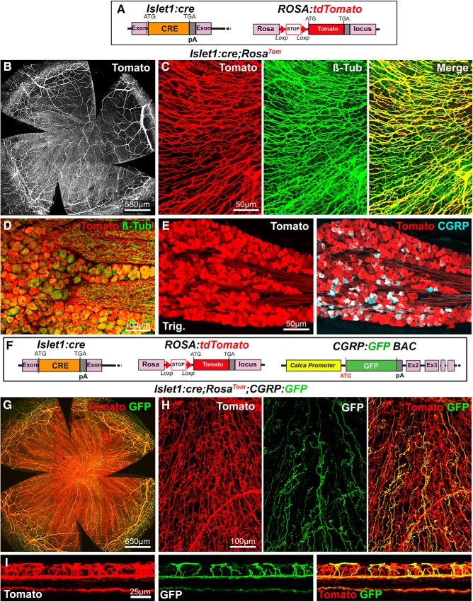
Figure 4.
Visualization of corneal axons in Islet1:cre adult mice. B, C, G, H, Maximal intensity z-projection confocal stacks from adult whole-mount corneas. A, Description of the mouse lines. RosaTom (see Fig. 2). In Islet1:cre knockin mice, the coding sequence of Cre was inserted in the isl1 gene by homologous recombination. B, C, In Islet1:cre;RosaTom mice, all corneal axons express Tomato (red). βIII-tubulin-immunoreactive axons (green) are also Tomato+ (see merge). D, E, Confocal images of cryostat sections of trigeminal ganglia. D, Colocalization of the GFP signal (green) and βIII-Tub immunoreactivity (red) in trigeminal neurons. E, All CGRP+ neurons (cyan) coexpress Tomato. F, Description of the mouse lines. Islet1:cre (see above). CGRP:GFP (see Fig. 1). RosaTom (see Fig. 2). G, GFP and Tomato expression in whole-mount cornea from an Islet1:cre;RosaTom;CGRP:GFP mouse. H, High magnification showing that Tomato (red) is expressed both by peptidergic (GFP+, green) and nonpeptidergic (GFP−) axons. I, A reslice of the cornea (54-μm-thick optical section) showing the location of the fluorescent axons. Yellow represents GFP+/Tomato+ peptidergic nociceptor axons. Red represents nonpetidergic nociceptor axons.

