Figure 5.
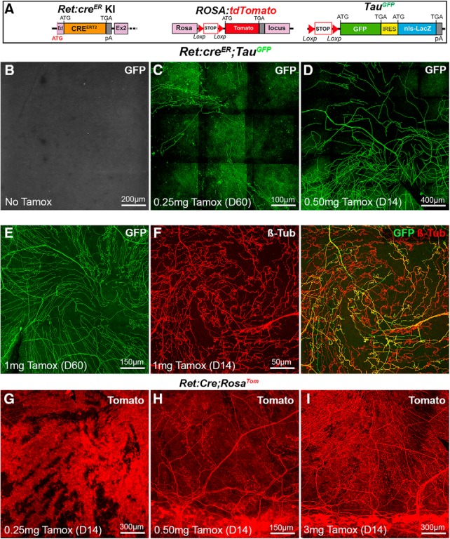
Visualization of corneal axons in Ret:creER adult mice. B–I, Maximal intensity z-projection confocal stacks from adult whole-mount corneas. A, Description of the mouse lines RosaTom and TauGFP (see Fig. 2). In Ret:creER knockin mice, the coding sequence of creERT2 was inserted in the first exon of the Ret gene by homologous recombination. B, In the absence of tamoxifen, no GFP signal is detected in the cornea of Ret:creER;TauGFPmice. C–E, The number of GFP+ axons increases with the dose of tamoxifen injected (0.25 mg-1 mg). Corneas were collected 14 d (D14) or 60 d (D60) after injection. F, Immunostaining for anti-βIII-tubulin shows that GFP is only expressed in a fraction of βIII-Tub+ corneal axons. G–I, Corneas from Ret:creER;RosaTom mice injected with increasing doses of tamoxifen injected (0.25–3 mg). At the lowest dose (G), many Tomato+ corneal cells are seen and mask Tomato+ axons. H, I, At higher doses, highly fluorescent cells are seen in the limbal region, and more Tomato+ axons are observed.

