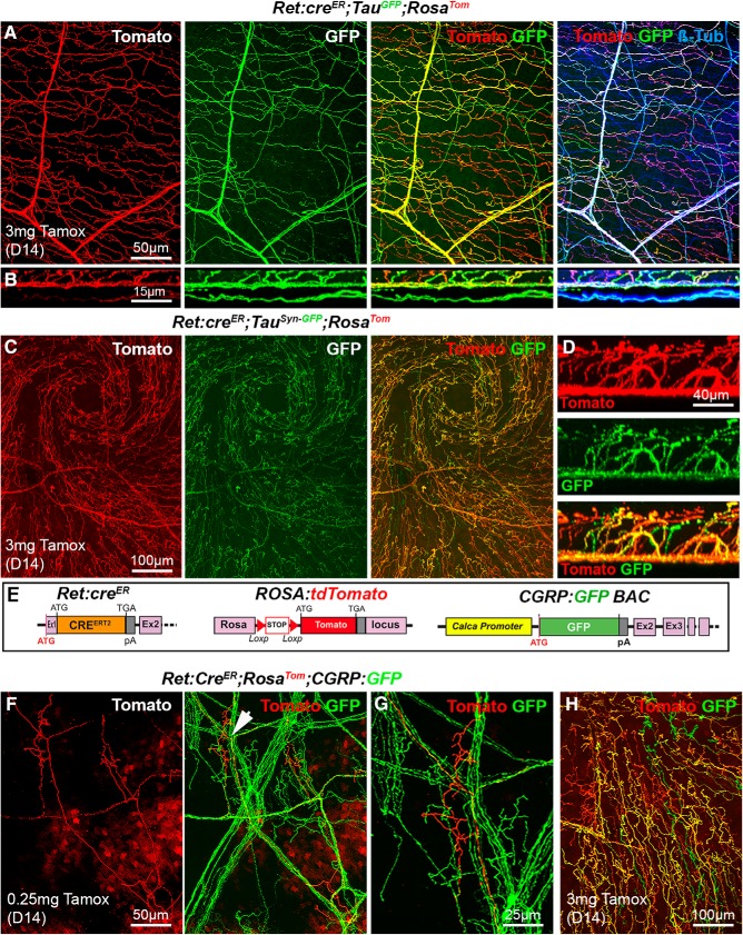
Figure 6.
Analysis of corneal nerves in Ret:creER compound mice. All images (except B and D) are maximal intensity z-projection confocal stacks from adult whole-mount corneas. A, Cornea from a Ret:creER;TauGFP;RosaTom mouse immunolabeled for βIII-tubulin. Some βIII-Tub+ axons (blue) also coexpress GFP and Tomato (and appear white). Other axons that only express GFP (right, green or cyan) or only Tomato (right, red or magenta). B, A reslice of the cornea (54-μm-thick optical section) illustrating the distribution of the fluorescent axons in the stroma and epithelium. C, Image of the apex of the cornea and axonal whorl from a Ret:creER;TauSyn-GFP;RosaTom mouse. The three types of axons are seen: GFP+, Tomato+, and a majority of GFP+/Tomato+ axons. D, A reslice of the cornea (54-μm-thick optical section). E, Description of the mouse lines. CGRP:GFP (see Fig. 1). RosaTom (see Fig. 2). Ret:creER (see Fig. 5). F, G, With a low dose of tamoxifen (0.25 mg), only a few Tomato+ axons and do not always overlap with GFP+ nociceptive axons. Middle, Arrowhead indicates the area seen on the high-magnification image of a single tomato+ terminal arbor (right). H, With a high dose of tamoxifen, most axons coexpress GFP and Tomato, but a few only express a single fluorescent protein.

