Figure 4.
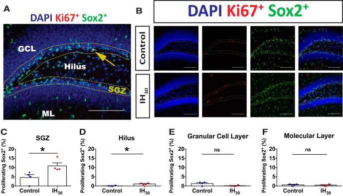
IH30 stimulates region-specific SOX2+ cell proliferation in the dentate gyrus. A, Representative section of dentate gyrus stained for Sox2 (green), Ki67 (red), and DAPI (blue). SGZ is outlined by yellow dotted lines. Counts were performed in the ML, GCL, SGZ, and hilus. The yellow arrow indicates a Ki67+/Sox2+ double-positive cell residing within the SGZ. Scale bar, 100 μm. B, Representative images of Ki67 and Sox2+ labeling in control (top) and IH30 (bottom) animals. SGZ is outlined in yellow. Blue channel depicts DAPI-labeled nuclei on left, red channel depicts Ki67+ labeling second from left, green channel depicts Sox2+ labeling second from right, and a merge is on the right. Scale bars, 100 μm. C–F, Quantified proportions of double-labeled Sox2+/Ki67+ cells (control, n = 4; IH30, n = 4) in the SGZ (t(5.993) = 2.747, p = 0.034), hilus (t(4.415) = 4.775, p = 0.0069), GCL (t(3.580) = 2.414, p = 0.0808), and ML (t(5.89) = 0.4592, p = 0.6625). *p < 0.05.

