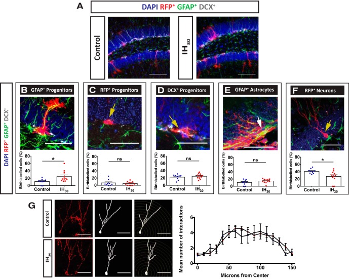
Figure 6.
IH30 exposure alters neural progenitor cell fate within the dentate gyrus. A, Representative images of tissue stained for birth-labeled RFP+ cells (red), GFAP (green), DCX (gray), and DAPI (blue) from control (left) and IH30-exposed (right) mice. Scale bars, 100 μm. B–F, The proportion of: RFP+/GFAP+-colabeled cells with radial glial morphology were significantly different between groups (control, n = 9; IH30, n = 11; t(15.16) = 2.635, p = 0.0186; B); RFP+ neural progenitor cells in the SGZ were unchanged between groups (control, n = 8; IH30, n = 11; t(12.60) = 0.4915, p = 0.6315; C); RFP+/DCX+-colabeled progenitor cells were unchanged between groups (control, n = 8; IH30, n = 11; t(17.13) = 0.7422, p = 0.4680; D); RFP+/GFAP+-colabeled cells with astrocytic morphology were not significantly different between the two groups (control, n = 8; IH30, n = 11; t(14.68) = 1.267, p = 0.2250; E); RFP+ cells that exhibit neuronal morphology were reduced in IH30 mice (control, n = 8; IH30, n = 11; t(13.97) = 2.730, p = 0.0163; F). Scale bars: B–F, 50 μm. G, Sholl analysis revealed that there were no significant changes in morphology as characterized by the number of intersections in dendritic arborization (control, n = 7; IH30, n = 10; F(30,320) = 0.750, p = 0.828). Representative images of neurons from control (top) and IH30 (bottom) used for analysis on left. Scale bars: 50 μm. *p < 0.05.

