Figure 2.
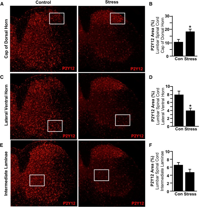
RSD caused region-specific microglial activation in the spinal cord. Male C57BL/6 mice were subjected to 6 d of RSD (Stress) or left undisturbed as controls (Con). Mice were perfused, and spinal cords were PFA fixed 14 h after the last day of stress. Microglial activation (P2Y12 expression) was assessed in the lumbar spinal cord. A, Representative images within the cap of the dorsal horn of P2Y12 labeling. B, P2Y12 proportional area was determined in the cap of the dorsal horn. C, Representative images within the lateral ventral horn of P2Y12 labeling. D, P2Y12 proportional area was determined in the lateral ventral horn. E, Representative images within the intermediate laminae of P2Y12 labeling. F, P2Y12 proportional area was determined in the intermediate laminae. Boxed insets represent location where proportional area was measured. Error bars indicate mean ± SEM. *Significantly different from control mice (p < 0.05; F-protected after analysis).

