Figure 4.
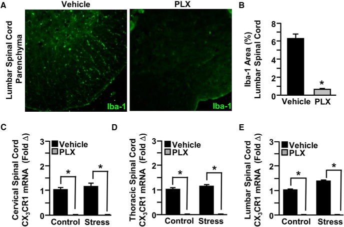
CSF1R antagonist PLX5622 depleted microglia in the spinal cord. Male C57BL/6 mice were provided ad libitum diets of PLX5622 (PLX, 1200 ppm chow) or vehicle (Veh) chow for 14 d. Mice were perfused, and spinal cords were PFA fixed to determine microglial ablation. Microglial activation (Iba-1) was assessed in the lumbar spinal cord parenchyma. A, Representative images within the lumbar spinal cord parenchyma of Iba-1 labeling. B, Iba-1 proportional area was determined in the lumbar spinal cord parenchyma. A separate set of mice provided with PLX5622 or vehicle chow for 14 d were then exposed to RSD (Stress) or left undisturbed as controls. Fourteen hours after the last day of stress, spinal cords were collected and dissected for mRNA analyses. CX3CR1 mRNA levels were determined in the (C) cervical, (D) thoracic, and (E) lumbar spinal cord. Error bars indicate mean ± SEM. *Significantly different from control mice (p < 0.05; F-protected after analysis).

