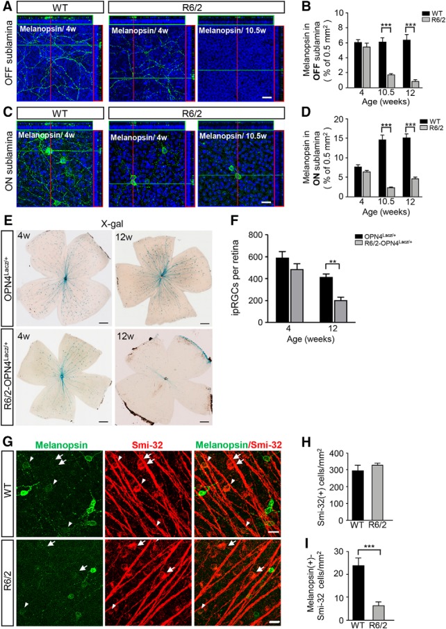
Figure 2.
Alteration of M1-ipRGCs and non-M1 ipRGCs in R6/2 mice at the symptomatic stage. A, C, Representative confocal images showing the changes of melanopsin-labeled processes in the OFF (A) and ON (C) sublaminae of the IPL in 4- and 10.5-week-old R6/2 mice compared with control mice at the indicated ages. B, D, Distribution of melanopsin in the OFF (B) and ON (D) sublaminae of IPL in the indicated mice at the different ages were measured by Fiji software and the expression levels of somatic and dendritic melanopsin relative to a retinal area (0.5 mm2) are presented; in an individual mouse, 6 retinal areas were randomly selected for the quantification (n = 3 for each group). E, X-gal-labeled ipRGCs in the retinas of OPN4Lacz/+ and R6/2-OPN4Lacz/+ mice at the ages of 4 and 12 weeks are shown. F, Bar graph presenting total X-gal-labeled M1-ipRGCs in R6/2-OPN4Lacz/+ (4 weeks, n = 3; 12 weeks, n = 3) and OPN4Lacz/+ mice (4 weeks, n = 5; 12 weeks, n = 4). G, Representative images showing melanopsin-positive ipRGCs with smi-32 immunoreactivity (arrowheads) and the smi-32-positive, melanopsin-negative RGCs (arrows) in the indicated mice at the age of 12 weeks. H, Numbers of Smi-32(+) cells were calculated in R6/2 and control retinas, n = 3 for each group. I, Numbers of cells immunoreactive for melanopsin/smi-32 in R6/2 retinas were lower than in control mice (n = 3 for each group). Scale bars: A, C, G, 20 μm; E, 500 μm. Data are presented as the means ± SEM. **p < 0.01, ***p < 0.001, two-way ANOVA and unpaired t test. ON sublamina, bottom part of the IPL; OFF sublamina, upper part of the IPL.

