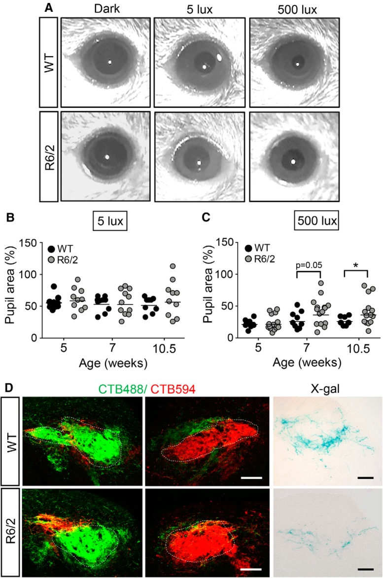
Figure 9.
Changes in pupil constriction in response to light in R6/2 mice. A, Pupil constriction induced by 30 s of light analyzed in R6/2 mice (n = 15) and control mice (n = 9) at the ages of 5, 7, and 10.5 weeks. Representative images show the pupillary constriction of 10.5-week-old mice in response to 5 and 500 lux light, respectively. B, C, Maximal response of pupil constriction extracted from 11 to 23 s during the 30 s constriction in response to light presented as the relative change in the pupil area. Note that 100% of the relative pupil area is defined as a pupil without constriction. Light-induced pupil constriction at 5 lux (B) and 500 lux (C) in the indicated mice is shown. D, Confocal images showing that double CTb-labeled RGC innervation in the bilateral OPN of the indicated mice at 12 weeks of age (n = 5–6 for each group) and the X-gal-labeled M1-ipRGC innervation in the OPN of R6/2-OPN4Lacz/+ or OPN4Lacz/+ mice at 12 weeks of age (n = 3–4 for each group). Data are presented as the means ± SEM. *p < 0.05, two-way ANOVA.

