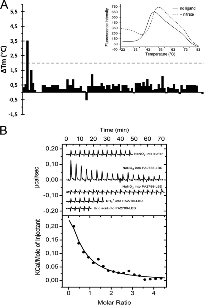
FIG 1.

Identification of nitrate as a PA2788-LBD ligand. (A) Thermal shift assays using compounds of Biolog compound array PM3B. Shown are the Tm changes with respect to the ligand-free protein. The insert shows the thermal unfolding curves of ligand-free PA2788-LBD and in the presence of nitrate. (B) Microcalorimetric binding studies of PA2788-LBD. The upper panel shows the heat changes caused by the injection of 2 mM (12.8-µl aliquots) NaNO3 into buffer and 36 µM PA2788-LBD as well as the titration of PA2788-LBD with 2 mM NaNO2, 2 mM ammonia, and 1 mM uric acid. The lower panel depicts the concentration-normalized and dilution heat-corrected integrated peak areas of the PA2788-LBD titration data with NaNO3. The line corresponds to the best fit using the “One binding site model” of the MicroCal version of ORIGIN.
