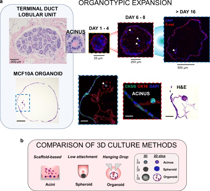
Fig. 1. Organotypic expansion in MCF10A cells cultured in 3D hanging drop system.
a TOP PANEL: representative image of a normal terminal duct lobular unit (TDLU) in a human breast tissue sample (University of Michigan IRB HUM00050330) and inset showing an acinar structure comparing with organoid formation through 16 days, with expression of epithelial marker E-cadherin and hollow lumen formation. White arrows indicate 3D acinar structures within organoids. BOTTOM PANEL: 3D MCF10A organoid after 16 days by H&E showing a large hollow structure (scale bar = 200 μm), blue inset at 20X showing internal features (scale bar = 50 μm), red inset at 60X magnification of a developing acinar structure (scale bar = 15 μm) (confocal images: CK5/6 = green, CK18 = red, nuclei = DAPI) and corresponding H&E image (scale bar = 15 μm). b diagram of conventional scaffold-based Matrigel culture or U-bottom spheroid formation compared with the hanging drop system. Legend shows characteristic sizes of acini, spheroid or organoid structures grown in 3D

