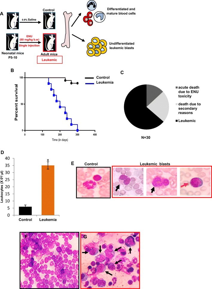
Fig. 1. Pathological features of ENU induced experimental leukemic mice.
a Outline of experimental strategy to induce leukemia in 5–10 days old Swiss albino litter pups by single intraperitoneal injection of ethyl-nitrosourea (ENU) at dose rate of 80 mg/kg b.w. b Kaplan-meier survival plot of ENU-induced leukemic mice (N = 18; median survival = 200 days). c Pie chart represented the percentage of mice (63.66%) developed leukemia after ENU induction. Rest of the mice died due to acute ENU toxicity (13.13%) and due to secondary reasons (23.21%). d The total leukocyte count of control and leukemic mice. Leukemic peripheral blood showed significant leukocytosis [*P < 0.05] (e) Representative control and leukemic blood films. Neutrophil and lymphocyte in control blood. Morphologically distinct leukemic blasts such as large cells with irregular nuclei indicated myeloblasts (black arrow) and small cells with round nuclei indicated lymphoblasts (orange arrow) [Magnification 1000X]. Representative bone marrow (BM) smears of (f) control and (g) leukemic mice. Leukemia BM showed accumulation of undifferentiated leukemic blasts (black arrows) [Magnification 400X]

