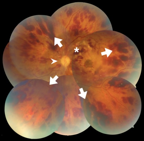
Figure 1. Montage image of the left retina shows optic disc pallor (arrowhead). Extensive superficial retinal hemorrhages are seen (arrows) and the retinal veins are dilated and tortuous consistent with central retinal vein occlusion. Few cotton wool spots are seen superotemporal to the optic disc (asterisk). Macular edema can also be made out.

