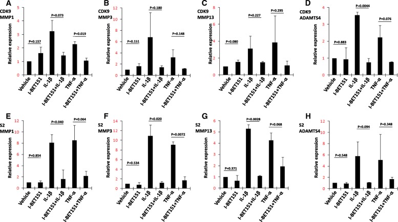
Fig. 6.
Identification of H4 acetylation after stimulation by IL-1β or TNF-α. a–c SW1353 cells were stimulated with or without IL-1β (10 ng/ml) or TNF-α (10 ng/ml) for 6 h and analyzed by ChIP for H4K5Ac, H4K8Ac, and H4K12Ac. The quantitative analysis of targeted promoter regions was determined by real-time PCR using specific primers for MMP1, MMP3, MMP13, and ADAMTS4. Relative fold-change values were calculated in comparison with the vehicle control that was set to 1 (n = 2)

