Figure 1.

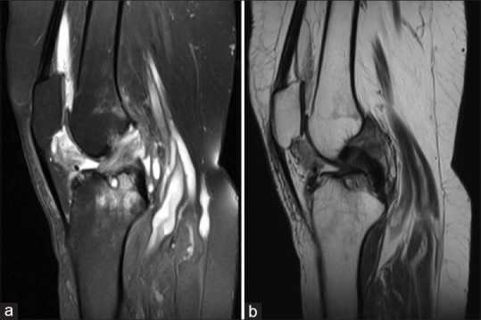
Sagittal view of the right knee magnetic resonance imaging; T2 view showing high-signal intensity in the anterior cruciate ligaments, Baker's cyst and absence of pigmented villonodular synovitis features (a and b)

Sagittal view of the right knee magnetic resonance imaging; T2 view showing high-signal intensity in the anterior cruciate ligaments, Baker's cyst and absence of pigmented villonodular synovitis features (a and b)