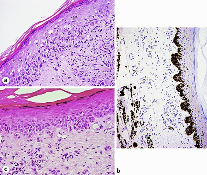
Fig. 2.
a Atypical melanocyte infiltrate in the superficial dermis. Pagetoid spreading is also identified. H&E. ×40. b A dramatic increase of melanocytes is noted at the dermo-epidermal junction. Enlarged, atypical melanocytes are immunohistochemically identified in the mid dermis. Melan-A, ×20. c The periphery of the lesion shows the presence of nests of atypical melanocytes at the dermo-epidermal junction, in keeping with in situ melanoma.

