Figure 1.

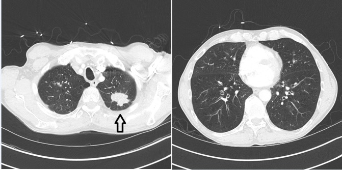
CT chest at time of diagnosis of lung cancer, including images of lower lungs at time of diagnosis; a spiculated 3.6 × 2.8 cm left upper lobe mass is demonstrated (black arrow).

CT chest at time of diagnosis of lung cancer, including images of lower lungs at time of diagnosis; a spiculated 3.6 × 2.8 cm left upper lobe mass is demonstrated (black arrow).