
Figure 2.
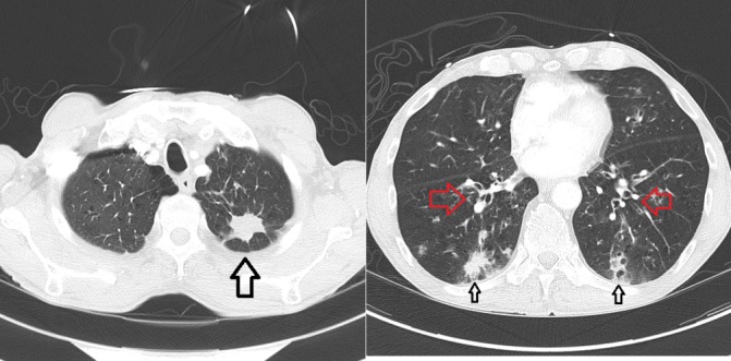
CT chest at time of diagnosis of allergic bronchopulmonary aspergillosis, 4 months into treatment; primary lung lesion in left upper lobe now 2.8 cm in length is decreased in size (large black arrow); new peribronchial thickening is noted (red arrows) and peripheral infiltrates (small black arrows).