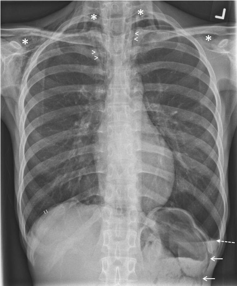
Figure 1.
Erect chest radiograph demonstrating a left-sided hydropneumothorax (dashed arrow, with air–fluid level seen), pneumomediastinum (arrowheads), subcutaneous emphysema (asterisk), pneumoperitoneum (short lines) and suggestion of pneumoretroperitoneum (solid arrow). No gross pulmonary lesion was evident.

