Fig. 3.
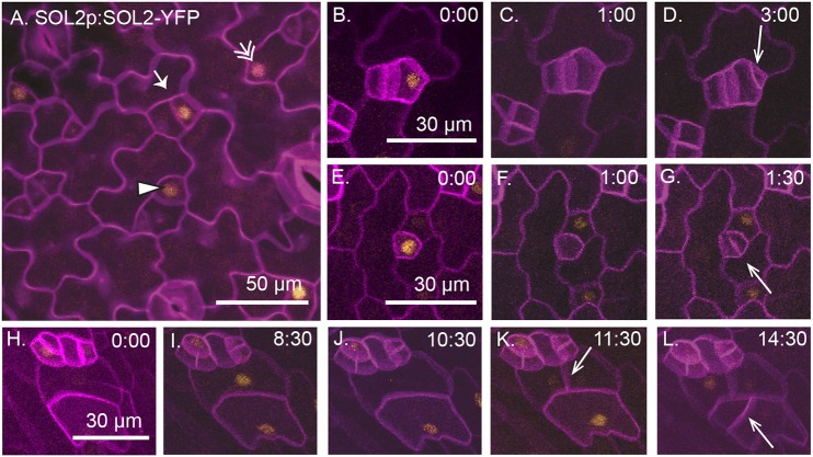
SOL2 is expressed in meristemoids, GMCs and pavement cells in a cell cycle-dependent manner. (A) A functional SOL2-YFP reporter is expressed in meristemoids (arrow), GMCs (arrowhead) and pavement cells (double arrow) in 3 dpg abaxial cotyledon (full genotype: SOL2p:SOL2-YFP; sol1 sol2). Cell outlines are stained using propidium iodide (purple). (B-L) Time-lapse images of SOL1p:SOL1-YFP (yellow) with cell outlines marked using ML1p:RCI12A-mCherry (purple) in a wild-type background; time in h:min is noted in the top right of each image. Arrows indicate new cell divisions. (B-D) A meristemoid divides asymmetrically (arrow). (E-G) A GMC divides symmetrically (arrow). (H-L) Pavement cells divide (arrows). In each division, SOL2 expression disappears 1-2 h before cell division.

