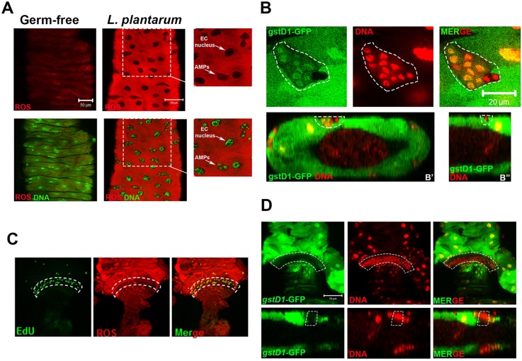
Fig. 1.
Discrete localization of cellular ROS within the Drosophila midgut and hindgut stem cell microenvironment. (A) ROS generation in the midgut ISC niche of germ-free third instar larvae colonized with L. plantarum for 4 h. Food also contained 100 μM of the ROS-sensitive hydro-Cy3 dye. Magnified images of boxed areas shown on right. (B) Detection of GFP from the ROS-responsive gstD1-gfp element in the midgut ISC niche of germ-free third instar larvae colonized with L. plantarum. (B′) Transverse section and (B″) longitudinal section of the larval midgut. (C) Detection of ROS using the hydro-Cy3 dye, and of EdU-positive cells in the hindgut pylorus of w1118 larvae that were colonized with L. plantarum for 2 h. (D) Detection of GFP using gstD1-gfp in the hindgut pylorus of germ-free third instar larvae that were colonized with L. plantarum 4 h. Bottom panels show longitudinal section of the hindgut pylorus. Region of low GFP is outlined by dashed white lines. Dashed white lines outline the midgut stem cell niche (B) and the hindgut proliferation zone (C,D). Scale bars: 50 μm in A,D; 20 μm in B.

