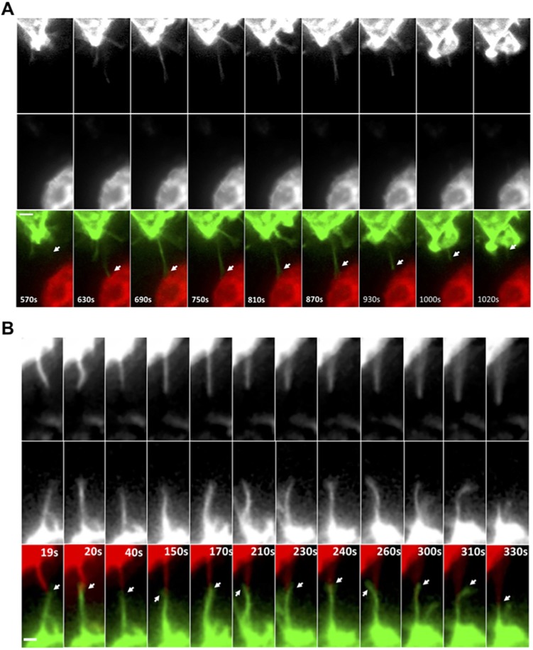
Fig. 3.
Live-cell imaging of heterotypic TNT formation between tumor cells and macrophages. (A) Time-lapse imaging of GFP–CAAX RAW/LR5 macrophages in co-culture with mCherry–CAAX MTLn3 tumor cells showing TNT-like protrusion initiated from macrophage towards a tumor cell (white arrows, lower panels). Upper and middle panels show individual channels. See Movies 5 and 6. (B) Time-lapse imaging of GFP–CAAX RAW/LR5 macrophages in co-culture with mCherry–CAAX MTLn3 tumor cells showing TNT-like protrusions extending from both cell types where they appear to intertwine and/or interact at the tip of the TNT-like protrusion (white arrows, lower panels). See Movie 7. Duration of original sequence at least 30 min. Magnification 60×, 2×2 binning. Frame interval: 10 s. Scale bars: 2 μm.

