Fig. 7.
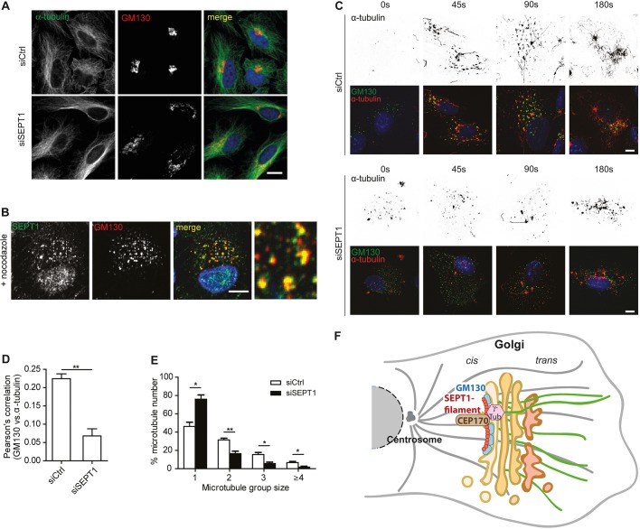
SEPT1 facilitates microtubule nucleation at the Golgi. (A) Staining of α-tubulin and GM130 in control and in SEPT1-depleted cells. (B) SEPT1 localization to GM130-positive Golgi objects in RPE1 cells is not affected by nocodazole administration. Scale bar: 10 µm. (C) GM130- and α-tubulin staining in control and SEPT1-depleted RPE1 cells, fixed at the indicated time points after nocodazole washout. Scale bar: 10 µm. (D) The Pearson's correlation coefficient of α-tubulin and GM130 staining decreases upon depletion of SEPT1, demonstrating that α-tubulin becomes less abundant at early Golgi membranes. Data are represented as mean±s.e.m., unpaired two-tailed Student's t-test, **P<0.01 (N=3; siScr: n=176; siSEPT1: n=96; t=6.613; d.f.=4). (E) Number of microtubules nucleated in 45 s upon nocodazole washout as determined for individual GM130-positive objects in control and SEPT1-depleted RPE1 cells (see also Fig. S7H). Data are represented as mean±s.e.m., unpaired two-tailed Student's t-test, **P<0.01, *P<0.05, (microtubule group size 1: t=4.478; d.f.=4; microtubule group size 2: t=4.652; d.f.=4; microtubule group size 3: t=3.65; d.f.=4; microtubule group size ≥4: t=3.085; d.f.=4). (F) Model for SEPT1 function at the Golgi. GM130 (in blue) recruits SEPT1 (in red) to cis-Golgi membranes. SEPT1 is organized in a filament-like fashion and serves as an anchor for the association of CEP170 (brown) and γ-tubulin (pink) to nucleate Golgi-derived microtubules (green).

