FIGURE 5.

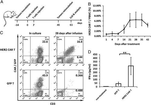
The expression and survival of engineered T cells in patient-derived xenograft model. A, Schematic diagram outlining the protocol of the experiment with the engraftment of the tumor tissue from patients to NPG mice. Mice were subcutaneously inoculated with dissected tumor masses from patients and infused with 5×10⁶ T cells on days 7 and 14. The P1 xenograft tumor tissues were retransferred on day 46 and excised on day 63 for tumor analysis. B, In vivo persistence of HER2-specific CAR-T cells. Variable expression levels of HER2-specific CAR-T cells were detected in the blood samples of mice. CAR+ cells were detected by GFP fluorescence in the blood of all mice after the infusion. HER2-specific CAR-T cells were detected every week after infusion. Error bars represent the SEM of 6 mice. C, Both GFP-T and HER2-specific CAR-T cells were detected by florescence-activated cell sorting analysis before and after T-cell infusion. HER2-specific T cells showed significant proliferation in tumor-bearing mice. D, In vivo, HER2-specific CAR-T cells may produce IFN-γ. IFN-γ may be detected in mice peripheral blood 7 days after infusion. Error bars represent the SEM of 6 mice. **P<0.005. CAR-T indicates chimeric antigen receptor T cell; HER, human epidermal growth factor receptor; IFN, interferon; PBMC, peripheral blood mononuclear cell; SEM, SE of mean.
