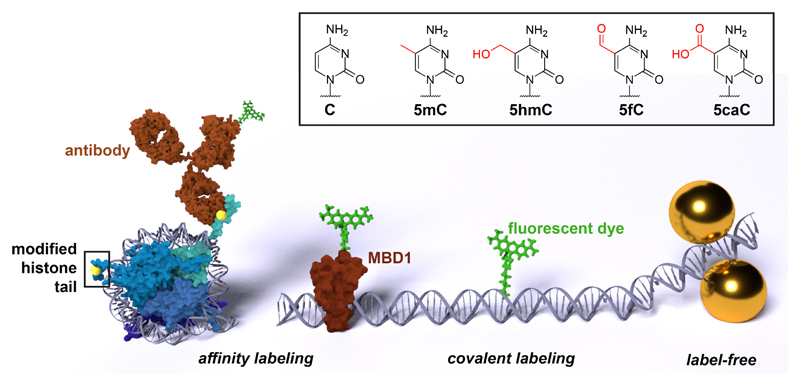
Figure 1.
The detection schemes discussed in this review: affinity labeling via fluorescently-labeled antibodies or other binding proteins such as the methyl binding domain protein 1 (MBD1), direct covalent labeling with a fluorescent dye, and the label-free detection via surface-enhanced Raman scattering (left to right, sizes not to scale). The chemical structure of cytosine (C), 5-methylcytosine (5mC), 5-hydroxymethylcytosine (5hmC), 5-formylcytosine (5fC), and 5-carboxycytosine (5caC) is shown in the box.

