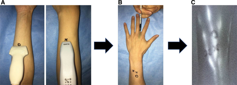
Fig. 1.

A, The identified lymphatic vessels using UHFUS were marked as cross with a black pen, and ones using CHFUS were marked as circle with a black pen. B, After the identification of lymphatic vessels using ultrasound, ICG was injected subcutaneously into the extremity. C, Then, the marked points were checked with ICG lymphography.
