Fig. 5.
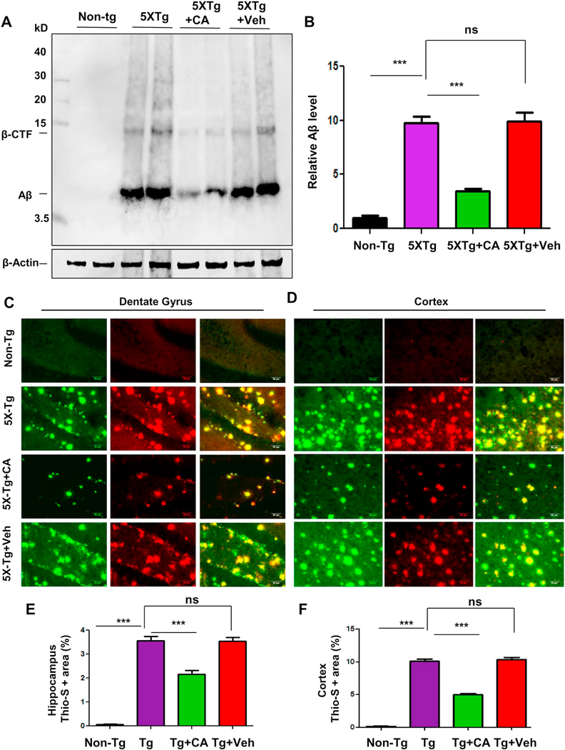
Administration of cinnamic acid reduces amyloid beta plaque deposition in the 5×FAD model of AD. Six months old 5×FAD mice (n = 8/group) were treated via oral gavage with cinnamic acid (100 mg/kg/day) or vehicle (0.5% methylcellulose) daily for one month following which they were sacrificed and (A-B) the cerebral amyloid-beta levels were analyzed by (A) immunoblot analysis of hippocampal homogenates using Aβ 6E10 monoclonal antibody and (B) densitometric analysis of relative Aβ (Aβ/Actin) levels with respect to non-transgenic; (C–F) Cerebral amyloid plaque deposition was monitored by colabeling free floating hippocampal sections with thioflavin-S and Aβ 6E10 antibody. Thio-S and Aβ positive plaques in (C) Dentate gyrus regions of the hippocampus and (D) Cortex are shown. Quantification of the thio-s area fraction (thio-S positive area represented as a percentage of total area) in the hippocampus (E) and cortex (F) was performed using ImageJ (analyze particle feature). All data represents mean ± SEM. All statistical analysis were performed by one way ANOVA followed by Tukey’s multiple comparison test; * p < .05; ** p < .01; *** p < .001.

