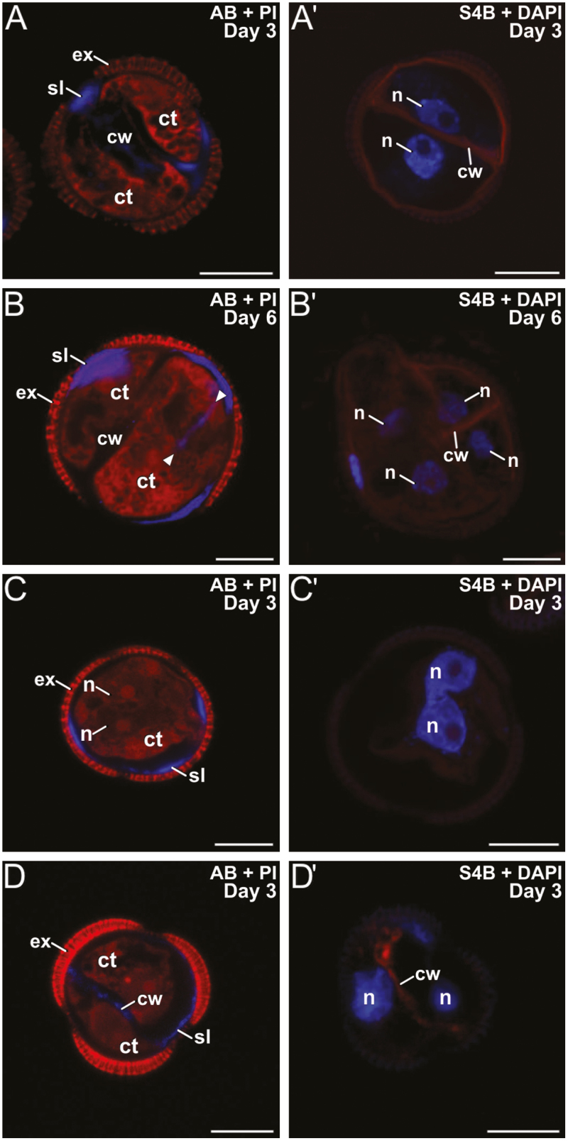
Fig. 5.
Microspores of rapeseed line DH4079 treated with caffeine. Paired images are shown of staining with aniline blue (AB) for callose and propidium iodide (PI) for cytoplasm, and with S4B for cellulose (red) and DAPI. Embryogenic microspores treated with 1 mM caffeine at (A, A′) 3 d old and (B, B′) 6 d old. The arrowheads indicate inner cell wall fragments. (C, C′) Embryogenic microspores treated with 10 mM caffeine at 3 d old. Note the presence of binucleated cells without inner walls and with two closely apposed (C) or even fused nuclei (C′). (D, D′): Examples of the few 3-d-old 2-celled embryogenic microspores treated with 10 mM caffeine that developed inner walls. ct, cytoplasm; cw, cell wall; ex, exine; n, nucleus; sl, subintinal layer. Scale bars are 10 µm.

