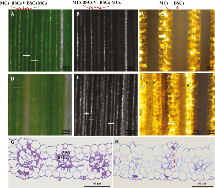
Fig. 2.
Abnormal leaf vein arrangement and asymmetric cell development of sistl1. (A, B) Fifth-leaf fragments of Yugu1 observed by optical (A) and confocal (B) microscopy. Arrows indicate the locations of mesophyll cells (MCs) and bundle sheath cells (BSCs) in one vascular bundle. White bars show the distance between two adjacent veins. (D, E) Fifth-leaf fragments of sistl1 observed by optical (D) and confocal (E) microscopy. (C, F) I2–KI-stained fifth-leaf fragments of Yugu1 (C) and sistl1 (F). Dark and light arrows indicate unstained BSCs and MCs. (G, H) Resin-embedded sections of Yugu1 (G) and sistl1 (H) fifth-leaf fragments. Triangles and squares denote abnormal BSCs and MCs in one vascular bundle.

