Fig. 3.
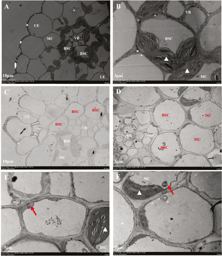
Reduced chloroplast biogenesis in sistl1. (A, B) Ultrastructure of Yugu1 fifth-leaf fragments observed by transmission electron microscopy (TEM) under ×0.5K (A) and ×1.5K (B) magnification. (C–F) Ultrastructure of fifth-leaf sistl1 fragments observed by TEM under ×0.5K (C), ×1.2K (D), ×2.0K (E) and ×2.5K (F) magnification. Normal cells in Yugu1 and sistl1 are labeled with white letters, with white triangles indicating normally developed chloroplasts in BSCs and MCs. Unusual empty cells in sistl1 are labeled with dark letters. Dark arrows indicate lysosome- or peroxisome-like organelles.

