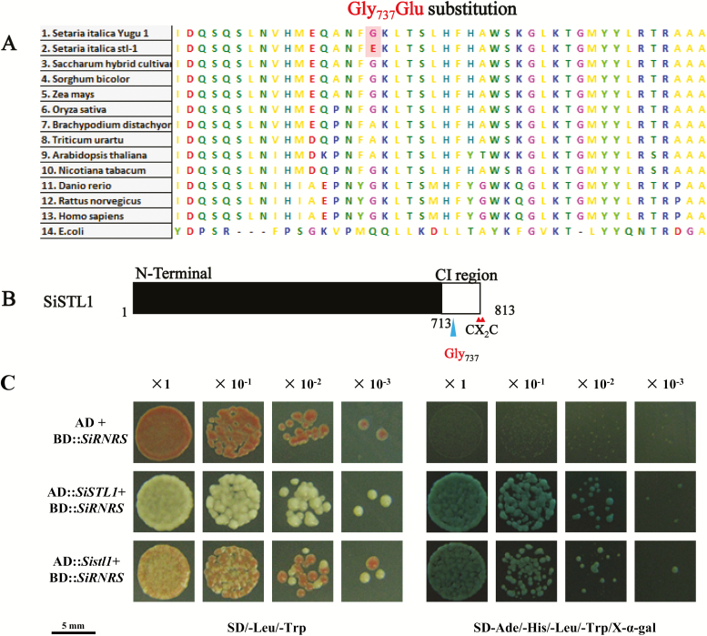
Fig. 7.
Sequence analysis of SiSTL1 and results of a yeast two-hybrid assay of SiSTL1 and SiRNRS. (A) Comparison of aligned sequences of SiSTL1 and homologous proteins from other species. (B) Schematic diagram of SiSTL1. Small triangles indicate the positions of the CX2C motif of SiSTL1. The last ~100 amino acids before the CX2C motif are designated as the C-terminal insertion (CI) region. The large triangle indicates the position of Gly737 in the CI region of SiSTL1. (C) Yeast two-hybrid analysis of SiSTL1 and SiRNRS. Dilutions are shown at the top (×10−1, yeast diluted 10 times; ×10−2, yeast diluted 100 times; ×10−3, yeast diluted 1000 times).

