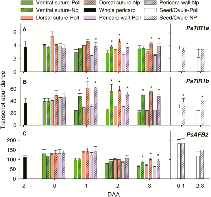
Fig. 4.
Relative transcript abundance of (A) PsTIR1a, (B) PsTIR1b, and (C) PsAFB2 in different tissues of pollinated (Poll) and non-pollinated (Np) fruits of pea. Pre-pollinated pericarps are from fruits at –2 d after anthesis (DAA). Pericarp tissues (ventral vascular suture, dorsal vascular suture, and pericarp wall) are from pollinated or non-pollinated fruits at 0–3 DAA. Seeds (fertilized ovules from pollinated fruits) and non-fertilized ovules (from non-pollinated fruits) are from fruits at 0–1 DAA and 2–3 DAA. Data are means (±SD), n=3 for pre-pollinated pericarps and pericarp wall tissue; n=3 for pericarp ventral and dorsal sutures, except for a few samples where n=2 due to the limited size of tissue that was available. At 3 DAA, non-pollinated ovaries were still green and turgid. Pairwise mean comparisons were made using a two-tailed Student’s t-test (P<0.05): * means for non-pollinated fruits are significantly different from those for pollinated fruits within a tissue, gene, and DAA (P<0.05).

