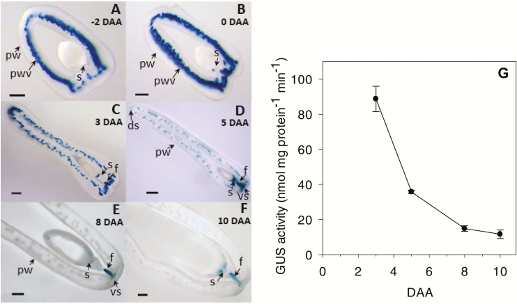
Fig. 5.
Developmental regulation of auxin activity in pea fruit. (A–F) Representative micrographs of GUS-stained fruit cross-sections and (G) GUS enzyme activity in the pericarp wall as detected by MUG assays in plants expressing the GUS gene under the regulation of the auxin-responsive DR5 promoter (DR5::GUS). The micrographs show DR5::GUS expression in (A) pre-pollinated fruit at –2 d after anthesis (DAA) and (B–F) in pollinated fruit at 0, 3, 5, 8, and 10 DAA. ds, dorsal vascular suture; f, funiculus; pw, pericarp wall, pwv, pericarp wall vasculature; s, proximal end of the seed; vs, ventral vascular suture. Scale bars: (A, B) 500 µm and (C–F) 1000 µm. GUS enzyme activity was quantified in the central pericarp wall of pollinated fruits at 3–10 DAA. Data are means (±SD), n=3.

