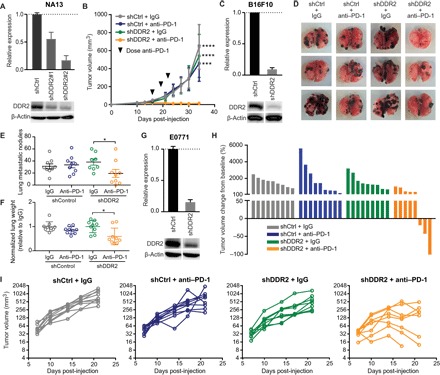
Fig. 2. In vivo evaluation of the therapeutic efficacy of targeting DDR2 combined with anti–PD-1 immunotherapy.

(A) Immunoblot of NA13 cells transduced with two different DDR2 shRNAs, with graph showing densitometric analysis of DDR2 protein levels. (B) Subcutaneous tumor growth in syngeneic mice receiving NA13 shDDR2 #1 cells stably expressing shControl (shCtrl) or shDDR2 (n = 4 to 5 mice per group). Mean ± SEM. ***P < 0.001, ****P < 0.0001. (C) Immunoblot of B16F10 cells with shControl or shDDR2 construct. (D) Representative images of murine pulmonary lung metastases at 22 days following intravenous (tail vein) inoculation of B16F10 cells. (E) Quantification of the number of metastatic B16F10 lung nodules (n = 9 mice per group). Mean ± SEM. *P < 0.05. (F) Lung weight of mice bearing B16F10 lung metastases (n = 9 mice per group). Mean ± SEM. *P < 0.05. (G) Immunoblot of E0771 cells with shControl or shDDR2 construct. (H) Waterfall plot showing change in E0771 mammary fat pad tumor volume compared to baseline before treatment. (I) E0771 mammary tumor volume as a function of time for each mouse. n = 8 to 9 mice per group.
