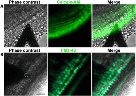
Fig. 3. Visualization of viable cochlear apical turn hair cells during noncontact FM-AFM measurements.

(A and B) Phase-contrast images showing the localization of the AFM cantilever with a 10-μm sphere (white dashed circle) over the OHCs. (A) Representative confocal image of the apical turn cells labeled with Calcein-AM confirming tissue viability. Scale bar, 45 μm. (B) Representative confocal image of the apical turn sensory epithelium transducing OHCs and IHCs labeled with 5 μM FM1-43, confirming hair cells’ proper MET channel functionality. Note that, with the 3-min incubation time used, cytoplasmic labeling of the sensory hair cells indicates that FM1-43 enters the cells mostly through their MET channels and not significantly through endocytosis (33). Scale bar, 25 μm. Images (A and B) were collected at the apical turn from different Strc+/−/Tecta−/− mice at P14.
