Figure 2.

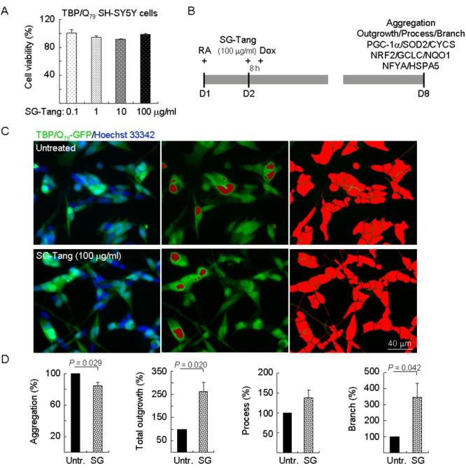
Neuroprotective effects of SG-Tang in TBP/Q79-GFP-expressing SH-SY5Y cells. (A) Cytotoxicity of SG-Tang (0.1−100 μg/ml) in uninduced cells using the MTT assay (n = 3). To normalize, the relative untreated cell viability was set as 100%. (B) Experimental flowchart. TBP/Q79-GFP SH-SY5Y cells were plated on dishes with retinoic acid (RA, 10 µM) added at day 1 to initiate neuronal differentiation. Next day, SG-Tang (100 μg/ml) was added to the cells for 8 h followed by inducing TBP/Q79-GFP expression (+ Dox, 5 µg/ml) for 6 days. Aggregation, neurite outgrowth, process, and branch were assessed by HCA. In addition, relative PGC-1α, NRF2, NFYA, and downstream targets were analysed by immunoblotting using specific antibodies. (C) Representative microscopic images of differentiated TBP/Q79-GFP-expressing SH-SY5Y cells untreated (top row) or treated with SG-Tang (bottom row), with nuclei counterstained in blue (left) or aggregates marked in red (middle). Shown right were images of the neurites and cell bodies outlined by red color for outgrowth quantification. (D) Relative aggregation, neuronal outgrowth, process, and branch of TBP/Q79-GFP-expressing SH-SY5Y cells with SG-Tang treatment (n = 3). To normalize, the relative aggregation, outgrowth, process, or branch level in untreated cells was set as 100%. P values: comparisons between treated and untreated cells.
