Figure 4.
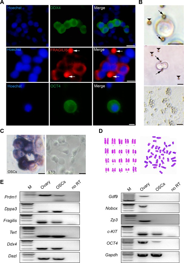
Morphology and characteristics of OSCs. (A) Immunofluorescence of DDX4 (green), FRAGILIS (red) and OCT4 (green) in mOSCs. Arrows: magnetic beads. Scale bars, 10 μm. (B) Examples of OSCs isolated with the Fragilis antibody and the associated pattern of OSCs with immunomagnetic beads. Arrows: magnetic beads associated with OSCs. Arrowheads: free beads. Scale bars, 10 μm, 10 μm, and 25 μm (From top to bottom). (C) Alkaline phosphatase staining for OSCs and STO. Scale bars, 10 μm. (D) Cytogenetic analysis showed that OSCs possessed a normal karyotype (40, XX). (E) Reverse transcription PCR analysis for the expression profile of OSCs using ovarian tissue as a positive control. M: 100 bp DNA marker; No RT, PCR of RNA sample without reverse transcriptase.

