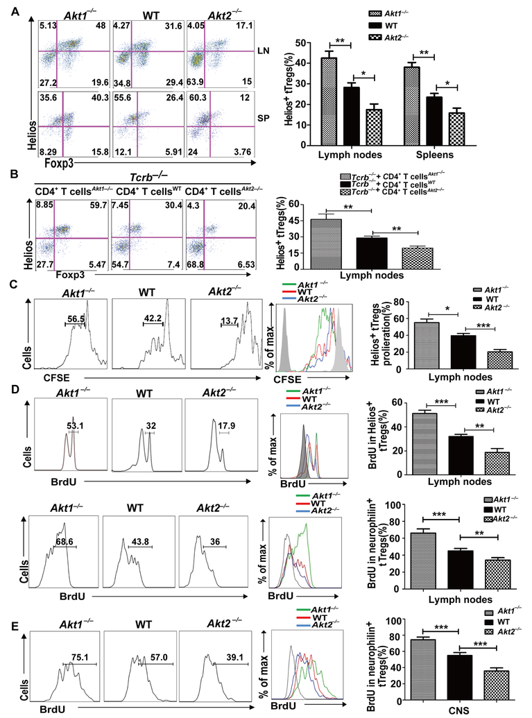
FIGURE 4.
Akt-1 inhibits, but Akt-2 facilitates, the proliferation of tTregs during EAE induction. (A) The spleen cells and lymph node cells from the mice (n = 5) described in Figure 2 were surface-stained with anti-CD4 and anti-CD25, and intracellularly stained with anti-Foxp3 and anti-Helios. *p < 0.05, **p < 0.01; Student t test. (B) 5 × 10⁶ CD4+ T cells from WT, Akt1–/–, and Akt2–/– mice were transferred by i.v. injection into Tcrb–/– mice (n = 5). After 30 days equilibration, the recipient mice were immunized with MOG35–55 in CFA. 10 days after immunization, the mice were sacrificed. The draining lymph node cells were surface-stained with anti-CD4 and anti-CD25, and intracellularly stained with anti-Foxp3 and Helios. **p < 0.01; Student t test. (C) WT, Akt1–/–, and Akt2–/– mice (n = 5) were immunized with MOG35–55 in CFA. At day 8, we collected the draining lymph node cells from each group, labeled with CFSE, and cultured them in the presence of 20 μg/ml MOG35–55 for three days. The cells were then surface-stained with anti-CD4 and anti-CD25, and intracellularly stained with anti-Helios and anti-Foxp3. The expression of Helios within CD4+CD25+Foxp3+ T cells was determined. *p < 0.05, ***p < 0.001; Student t test. (D and E) WT, Akt1–/–, and Akt2–/– mice (n = 5) were immunized with MOG35–55 in CFA. On day 5, the mice were injected with BrdU at 1 mg/mouse every 12 h for three consecutive days. The mice were sacrificed on day 8. The BrdU incorporation in CD4+CD25+Foxp3+Helios+ or CD4+CD25+Foxp3+neurophilin+ T cells in the draining lymph nodes (D) and CNS (E) of WT, Akt1–/–, and Akt2–/– mice was determined. **p < 0.01, ***p < 0.001; Student t test. The data shown are one representative of three independent experiments.

