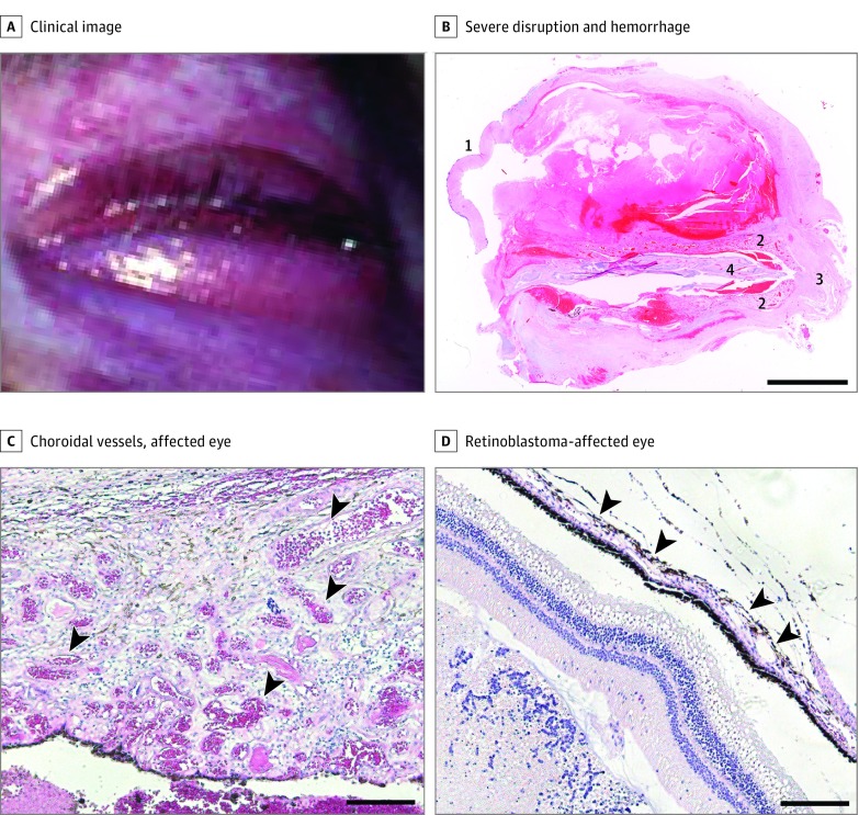
Figure 1. Patient With Sturge-Weber Syndrome and Choroidal Capillary Malformation.
A, Clinical image of the patient with choroidal capillary malformation showing diffuse capillary stain affecting the face and eye. B, Hemotoxylin-eosin–stained ocular tissue section showing severe disruption and hemorrhage. Major discernible structures are the cornea (1), the enlarged choroid (2), the optic nerve (3), and retinal tissue (4). Scale bars: 5 mm. C and D, Hemotoxylin-eosin–stained choroidal tissue sections. The affected eye is thickened and shows an abundance of blood-filled choroidal vessels (C, arrowheads) compared with the retinoblastoma-affected eye (D, where choroidal vessels are flat and close to the retinal pigment epithelium [arrowheads]). Scale bars: 200 μm.

