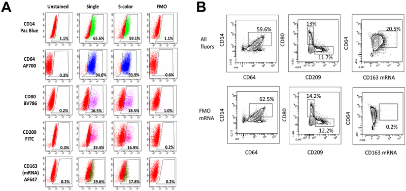
Figure 4. Several fluorophores are compatible with the use of CD163 mRNA Probe.
THP-1 cells were treated separately with M(LPS+IFNγ), M(IL-4), and M(IL-10) for 24 hours, then pooled together and processed as either unstained, stained with a single fluor, stained for all 5 indicated fluors or stained with all 5 fluors minus the indicated fluor (FMO, fluorescence minus one). (A) Percent of FSC vs SSC live, singlet population for each fluor is depicted in histogram form as SSC vs. Fluor. (B) Gating for subpopulations with all fluors included in staining or all fluors excluding AF647 CD163 mRNA probe (FMO mRNA). Results are representative of three separate experiments.

