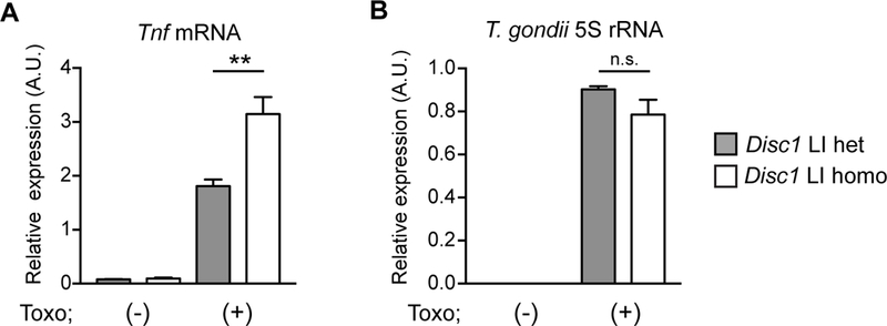
Figure 1: Increased anti-T. gondii immune response in primary cortical glial cells from mice with loss of function of Disc1 gene.

Primary cortical glial cells were prepared from postnatal day 2 (P2) pups of mating pairs of Disc1 LI heterozygous mice. Disc1 LI heterozygous and homozygous pups of the same litters were used for the study. On day 10, glial cells were infected with T. gondii tachyzoites and harvested at 48 h post-infection for qRT-PCR analysis. A. Enhanced induction of Tnf mRNA in Disc1 LI homozygous glial cells compared to Disc1 LI heterozygous glial cells. B. No difference in the amount of intracellular T. gondii between Disc1 LI mice and their heterozygous littermates. T. gondii 5S rRNA levels were measured by qRT-PCR in glial cell lysates to estimate the number of T. gondii. **p<0.01. n.s., not significant. Error bars show s.e.m.
