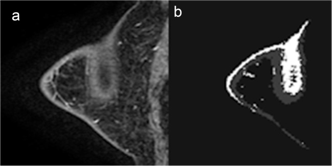
Fig. 5.
FGT and BPE segmentation. a Selected T1 sagittal postcontrast image. b Segmented FGT and BPE. MR acquisitions with relatively poor fat saturation along the subcutaneous skin margins. Incomplete fat saturation was combined with volume averaging in the out-of-plane direction resulting in apparent high signal intensity centrally within the breast tissue

