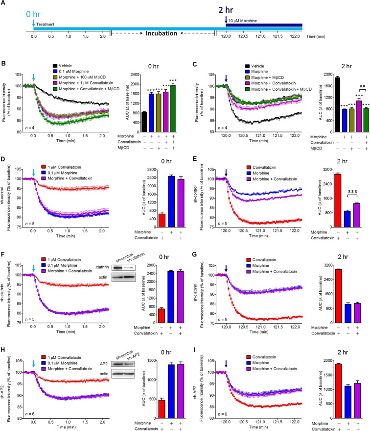
Figure 3.
Effects of convallatoxin on morphine-mediated membrane potential hyperpolarization in MOR-expressing AtT-20 cells. (A) Flowchart showing experiments testing the effects of convallatoxin on morphine activation of GIRK channels. AtT-20 Cells were transfected with myc-MOR expression plasmid for 24 h prior to all membrane potential assays. (B) Acute effects of each treatment in MOR-expressing AtT-20 cells were measured using membrane potential assay. AUC: F4,15 = 23.97, p < 0.0001 (1-way ANOVA). (C) After 2 h of incubation, chronic effects of each treatment on MOR desensitization were determined by rechallenging cells with 10 μM morphine. AUC: F4,15 = 84.32, p < 0.0001 (1-way ANOVA). (D,F,H) AtT-20 Cells were co-transfected with myc-MOR and sh-control (D), sh-clathrin (F), or sh-AP2 (H) for 24 h prior to membrane potential assay. Silencing of clathrin (F) and AP2 (H) did not attenuate the acute effect of convallatoxin. Immunoblot showing expression of clathrin (F) or AP2 (H) in clathrin- or AP2-knockdown AtT-20 cells (upper panel). AUC: (D) F2,12 = 88.35; (F) F2,12 = 323.2; (H) F2,15 = 75.45; all p < 0.001 (1-way ANOVA). (E, G, I) Both clathrin (I) and AP2 (G) were involved in the chronic convallatoxin effect. AUC: (E) F2,12 = 458.8; (G) F2,12 = 287.2; (I) F2,15 = 42.45; all p < 0.001 (1-way ANOVA). ***p < 0.001 versus vehicle group. ##p < 0.01 versus morphine + convallatoxin group. $$$p < 0.001 versus morphine-alone group (Newman-Keuls post hoc tests). All values indicate the mean ± SD.

